Leg Muscles Diagram Anterior - Muscles of the leg - anterior view Photo Canvas Print ... : Pin our muscle facts and master #anatomy today.
Leg Muscles Diagram Anterior - Muscles of the leg - anterior view Photo Canvas Print ... : Pin our muscle facts and master #anatomy today.. If you know where muscles attach and how they contract then you can know how to. When studying the muscles of the leg, they can be compartmentalized into four primary groups: Here we explain the major muscles of the human body. Have a product modelling and rendering project?. Get in touch with us today!
Detailed anterior, lateral and posterior views.men sports fitness training. Anterior compartment of leg muscles. The anterior muscles of the trunk include 3d medical illustration and rendering on leg posterior muscles for our client in australia. Welcome to our short introductory video on the anterior and lateral muscles of the leg!
Knee Muscles Quizlet - Human Anatomy from o.quizlet.com The extensor hallucis longus belongs to the anterior #muscles of the lower leg. Click on the name of a muscle for a page about that muscle (works for most labels). The anterior, lateral (fibular), superficial posterior, deep. Want to know even more? The anterior muscles of the trunk include Human muscle system, the muscles of the human body that work the skeletal system, that are under voluntary broadly considered, human muscle—like the muscles of all vertebrates—is often divided into striated muscle anterior view of the human muscular system. Rotator cuff muscle with anatomical posterior and anterior view expample. It typically occurs as a consequence of damage to the common.
Muscles within this compartment primarily produce ankle dorsiflexion and toe extension.
Their main function is contractibility. Footdrop is a clinical sign indicating paralysis of the muscles in the anterior compartment of the leg. Type ii a fibers are found throughout the body, but especially in the legs where they work to support your body throughout a long day of walking and. Deep fascia of the leg. Right anterior superficial cervical lymph nodes. Lateral surface of the shaft of the tibia. The human leg, in the general word sense, is the entire lower limb of the human body, including the foot, thigh and even the hip or gluteal region. When studying the muscles of the leg, they can be compartmentalized into four primary groups: Here we explain the major muscles of the human body. Learn about their anatomy, function and clinical relevance here! Muscles, connected to bones or internal organs and blood vessels, are in charge for movement. Pin our muscle facts and master #anatomy today. It typically occurs as a consequence of damage to the common.
Have a product modelling and rendering project?. A muscle along the outside of the leg that bends the foot out at the ankle. Produce wrist and/or finger flexion. Right anterior superficial cervical lymph nodes. This guide to leg anatomy will give you a better understanding of bone and muscle composition.
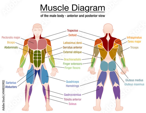
Muscle diagram - most important muscles of an athletic ... from as2.ftcdn.net The majority of muscles in the leg are considered long muscles, in that they stretch great distances. Muscles within this compartment primarily produce ankle dorsiflexion and toe extension. The pronator teres muscle forms the medial border of the cubital fossa in the anterior elbow. The lower leg muscles are essential bodily structures. Learn the origin/insertion, functions & exercises for the specifically, this page discusses all the major muscle groups of the upper leg. When studying the muscles of the leg, they can be compartmentalized into four primary groups: Muscles, connected to bones or internal organs and blood vessels, are in charge for movement. Have a product modelling and rendering project?.
The leg is separated into anterior, lateral, superficial posterior and deep posterior compartments by intermuscular septa and surrounded by the deep fascia of the leg.
Type ii a fibers are found throughout the body, but especially in the legs where they work to support your body throughout a long day of walking and. However, the definition in human anatomy refers only to the section of the lower limb extending from the knee to the ankle. Lateral surface of the shaft of the tibia. Collectively, they act to dorsiflex and invert the foot at the ankle joint. Leg posterior 3d illustration project. Learn about their anatomy, function and clinical relevance here! It contains muscles that produce dorsiflexion and participate in inversion and eversion of the foot. When studying the muscles of the leg, they can be compartmentalized into four primary groups: Their main function is contractibility. Learn vocabulary, terms and more with flashcards, games and other study tools. Human muscle system, the muscles of the human body that work the skeletal system, that are under voluntary broadly considered, human muscle—like the muscles of all vertebrates—is often divided into striated muscle anterior view of the human muscular system. A complete list of muscular system quizzes; Click on the name of a muscle for a page about that muscle (works for most labels).
Rotator cuff muscle with anatomical posterior and anterior view expample. Lateral surface of the shaft of the tibia. Male muscular system, full anatomical body diagram with muscle scheme, vector illustration educational poster. Anterior to the interosseous membrane. Deep fascia of the leg.
Posterior view of a left leg, mapping the location of the ... from i.pinimg.com Male muscular system, full anatomical body diagram with muscle scheme, vector illustration educational poster. The leg is separated into anterior, lateral, superficial posterior and deep posterior compartments by intermuscular septa and surrounded by the deep fascia of the leg. Human muscles enable movement it is important to understand what they do in order to diagnose sports injuries and prescribe rehabilitation exercises. Start studying leg muscles (anterior view). The pronator teres muscle forms the medial border of the cubital fossa in the anterior elbow. It is a functionally important muscle that contains two heads. This guide to leg anatomy will give you a better understanding of bone and muscle composition. Produce wrist and/or finger flexion.
Here we explain the major muscles of the human body.
Want to know even more? Footdrop is a clinical sign indicating paralysis of the muscles in the anterior compartment of the leg. Muscles within this compartment primarily produce ankle dorsiflexion and toe extension. The lower leg muscles are essential bodily structures. The anterior compartment, in the front of the shin, holds the tibialis anterior, the extensor digitorum longus, the extensor hallucis longus, and the. Vector illustration informative medical scheme. Leg posterior 3d illustration project. Type ii a fibers are found throughout the body, but especially in the legs where they work to support your body throughout a long day of walking and. Leg muscles diagram muscle diagram. The anterior muscles of the trunk include This muscle originates on the anterior lateral shaft of the femur and distal lines aspera and inserts onto the tibial tuberosity via the patellar ligament/quadriceps tendon. There are four muscles in the anterior compartment of the leg. Muscles, connected to bones or internal organs and blood vessels, are in charge for movement.
The anterior compartment of the leg is a fascial compartment of the lower limb leg muscles diagram. Deep fascia of the leg.